Burkitt's Lymphoma (BL)
You are here
Definition
Burkitt's lymphoma (BL) is a cancer of the lymphatic system (in particular, B lymphocytes). It is a highly aggressive lymphoma that is usually found in extranodal sites or presenting as an acute leukemia.
Sample Cases
Click here for instructions on how to download the free FCS Express Reader to view and manipulate the sample cases.
| Case Name (click on case name to open) |
Comments | Size |
| BL_1.fey | An atypical case of Burkitt's Lymphoma | 3.89 Mb |
Epidemiology
Currently Burkitt's lymphoma can be divided into three main clinical variants: the endemic, the sporadic and the immunodeficiency-associated variants.
- The endemic variant occurs in equatorial Africa. It is the most common malignancy of children in this area. Children affected with the disease often also had chronic malaria which is believed to have reduced resistance to the Epstein-Barr virus and allowed it to take hold. Disease characteristically involves the jaw or other facial bone, distal ileum, cecum, ovaries, kidney or the breast.
- The sporadic type of Burkitt lymphoma (also known as "non-African") is another form of non-Hodgkin lymphoma found outside of Africa. The tumor cells have a similar appearance to the cancer cells of classical African or endemic Burkitt lymphoma. Again it is believed that impaired immunity provides an opening for development of the Epstein-Barr virus. It accounts for 30-50% of childhood lymphoma. Jaw is less commonly involved, comparing with the endemic variant. Ileo-cecal region is the common site of involvement.
- Immunodeficiency-associated Burkitt lymphoma is usually associated with HIV infection or occurs in the setting of post-transplant patients who are taking immunosuppressive drugs. Actually, Burkitt lymphoma can be the initial manifestation of AIDS.
By morphology (i.e. microscopic appearance) or flow immunophenotype, it is almost impossible to differentiate these three clinical variants. Immunodeficiency-associated Burkitt lymphoma may demonstrate more plasmacytic appearance or more pleomorphism, but these features are not specific.
Possible causes
Epstein Barr virus (EBV) has been found to play a role in BL. The EBV genome is found in the majority of neoplastic BL cells. Genetic abnormalities (c-MYC) play a role in the pathogenesis of BL.
Morphology
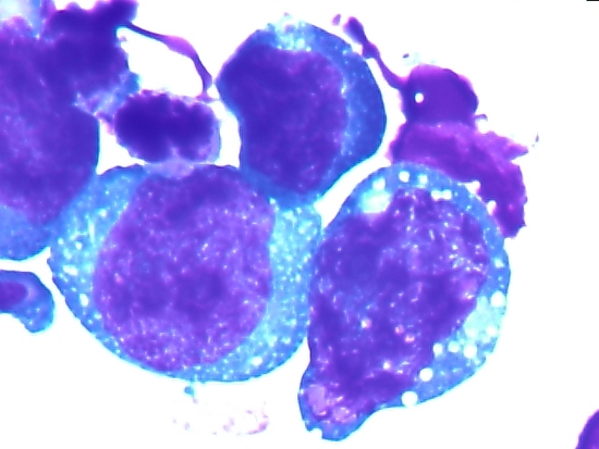
Consists of sheets of monotonous (i.e. similar in size and morphology) population of medium size lymphoid cells with highproliferative activity and apoptotic activity. The "starry sky" appearance seen under low power is due to scattered tingible-bodies laden macrophages (macrophages containing dead body of apoptotic tumor cells). The old descriptive term of "small non-cleaved cell" is misleading. The tumor cells are mostly medium in size (i.e. tumor nuclei size similar to that of histiocytes or endothelial cells). "Small non-cleaved cells" are compared to "large non-cleaved cells" of normal germinal center lymphocytes. Tumor cells possess small amount of basophilic cytoplasm. The cellular outline usually appears squared off.
 |
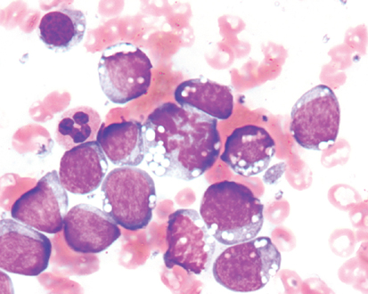 |
| Burkitt's lymphoma cells. Note lipid vacuoles and clear parachromatin. | Burkitt's lymphoma cells. Note lipid vacuoles and clear parachromatin. |
Immunophenotyping
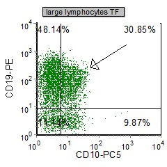
BL cells are bright CD45 and very large (high SSc and FSC). They can mimic large lymphocytes in a fresh tissue specimen (see below)
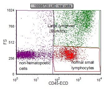 |
| Cells in BL can be initially identified using a SSC vs CD45 plot. These cells have moderate to high SSC/FSC and bright CD45. |
Phenotypic characteristics of BL are summarized in table below. Tumor cells are positive for CD45, the B cell markers CD19, CD22 and CD79a with a monoclonal light chain expression of kappa or lambda. CD10, CD38, CD43, CD71 and bcl-6 are also expressed. The blast markers CD34 and TdT are negative. SPF are very high (>30%) as is the proliferation marker Ki-67 (100%)
| Marker | Expression in BL |
| CD5 | - |
| CD10 | + |
| CD19 | +(mod) |
| CD20 | +(mod-bright) |
| CD22 | + |
| CD23 | -/(rare+) |
| CD25 | - |
| CD30 | - |
| CD43 | + |
| CD45 | + |
| CD79a | + |
| bcl-1 | - |
| bcl-2 | - |
| bcl-6 | + |
| Ki-67 | +(strong) |
| surface light chains (k or L) | + |
| TdT | - |
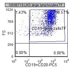
Example Dot Plots
Below are selected dual parameter dot plots that are useful in diagnosing BL.
 |
 |
 |
 |
 |
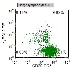 |
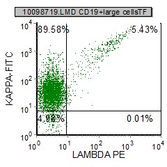
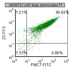
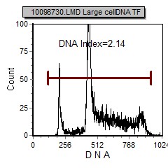
| In this case, 99% of the large CD19 B cells are selected and identify a monoclonal kappa light chain population | In this case, FMC7 and CD20 are expressed. An aneuploid population is identified (DI=2.14) with a very elevfated s-Phase fraction (high proliferation) | This case is negative for bcl-2, but CD10 is expressed. |
Other relevant tests
Cytochemistry: PAX-5 (B cell marker), Ki-67 (proliferation marker) is expected to be strongly positive. Many cases are EBER (EBV) positive.
Genetics: Burkitt lymphoma are associated with c-myc gene translocation. The most common variant is t(8;14)(q24;q32) while rarer variants include t(2;8)(p12;q24) and t(8;22)(q24;q11). A three-way translocation, t(8;14;18), has also been identified.[1] Burkitt's lymphoma are associated with c-myc gene translocation. The most common variant is t(8;14)(q24;q32) while rarer variants include t(2;8)(p12;q24) and t(8;22)(q24;q11). A three-way translocation, t(8;14;18), has also been identified.[1]
References
1. Liu D, Shimonov J, Primanneni S, Lai Y, Ahmed T, Seiter K (2007). "t(8;14;18): a 3-way chromosome translocation in two patients with Burkitt's lymphoma/leukemia". Mol. Cancer 6: 35. doi:10.1186/1476-4598-6-35. PMID 17547754.