Acute Lymphoblastic Leukemia (ALL) - General Information
You are here
Definition
Acute lymphoblastic leukemia (ALL), also known as acute lymphocytic leukemia, is a form of leukemia, or cancer of the white blood cells. Malignant, immature white blood cells continuously multiply and are overproduced in the bone marrow. ALL causes damage and death by crowding out normal cells in the bone marrow, and by spreading (metastasizing) to other organs. ALL is most common in childhood and young adulthood with a peak incidence at 4-5 years of age, and another peak in old age. The overall cure rate in children is 85%, and about 50% of adults have long-term disease-free survival. 'Acute' refers to the undifferentiated, immature state of the circulating lymphocytes ("blasts"), and to the rapid progression of disease, which can be fatal in weeks to months if left untreated.
Sample Cases
Click here for instructions on how to download the free FCS Express Reader to view and manipulate the sample cases.
| Case Name (click on case name to open) |
Comments | Size |
| ALL1 | A typical ALL case | 5.57Mb |
Epidemiology
The number of annual ALL cases in the US is roughly 4000, 3000 of which inflict children. ALL accounts for approximately 80 percent of all childhoodleukemia cases, making it the most common type of childhood cancer. It has a peak incident rate of 2-5 years old, decreasing in incidence with increasing age before increasing again at around 50 years old. ALL is slightly more common in males than females. There is an increased incidence in people with Down Syndrome, Fanconi anemia, Bloom syndrome, Ataxia telangiectasia, X-linked agammaglobulinemia and Severe combined immunodeficiency.
Possible causes
ALL is associated with exposure to radiation and chemicals in animals and humans. The association of radiation and leukemia in humans has been clearly established in studies of victims of the Chernobyl nuclear reactor and atom bombs in Hiroshima and Nagasaki. In animals, exposure to benzene and other chemicals can cause leukemia. Epidemiological studies have associated leukemia with workplace exposure to chemicals, but these studies are not as conclusive. Patients who are treated for other cancers with radiation and chemotherapy often develop leukemias as a result of that treatment.
Morphology
Morphologic subtyping of the various forms of ALL used to be done according to the French-American-British (FAB) classification,which was used for all acute leukemias (including acute myelogenous leukemia, AML). The FAB classification was:
- ALL-L1: small uniform cells
- ALL-L2: large varied cells
- ALL-L3: large varied cells with vacuoles (bubble-like features)
Note: The recent WHO International panel on ALL recommends that this classification be abandoned, since the morphological classification has no clinical or prognostic relevance. It instead advocates the use of the immunophenotypic classification mentioned below.
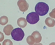
Example morphology in ALL.
 |
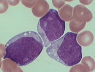 |
| Example of L1 blast | Example of L2 blast |
Immunophenotyping
Each subtype is then further classified by determining the surface markers of the abnormal lymphocytes, called immunophenotyping. There are 2 main immunologic types: pre-B cell and pre-T Cell. The mature B-cell ALL (L3) is now classified as Burkitt leukemia/lymphoma (BL). Subtyping helps determine the prognosis and most appropriate treatment in treating ALL.
Immunophenotypic characterisics of ALL:
| Marker | Blastic NK |
Precursor B-ALL | Precursor T-ALL |
| CD2 | - | - | + |
| CD3 | - | - | + |
| CD4 | + | - | +/- |
| CD7 | -/+ | - | +/- |
| CD10 | - | +/- | -/+ |
| CD11b | - | - | - |
| CD11c | - | - | - |
| CD13 | - | -/rare+ | - |
| CD14 | - | - | - |
| CD16 | - | - | - |
| CD19 | - | + | - |
| CD20 | - | -/rare+ | - |
| CD22 | - | +/- | - |
| CD33 | -/rare+ | -/rare+ | -/rare+ |
| CD34 | +/- | + | +/- |
| CD41 | - | - | - |
| CD45 | + | +/- | + |
| CD56 | + | - | -/+ |
| CD61 | - | - | - |
| CD64 | - | - | - |
| CD79a | - | + | - |
| CD117 | -/rare+ | - | -/rare+ |
| HLA DR | +(dim) | +/- | -/+ |
| TdT | -/+ | + | +/- |
| GPHA | - | - | - |
| cIgM | - | +/- | - |
Some cytogenetic subtypes have a worse prognosis than others. These include:
- A translocation between chromosomes 9 and 22, known as the Philadelphia chromosome, occurs in about 20% of adult and 5% in pediatric cases of ALL.
- A translocation between chromosomes 4 and 11 occurs in about 4% of cases and is most common in infants under 12 months.
Not all translocations of chromosomes carry a poorer prognosis. Some translocations are relatively favorable. For example, Hyperdiploidy (>50 chromosomes) is a good prognostic factor.