Follicular Lymphoma (FL)
You are here
Definition
Follicular lymphoma (FL) is the most common of the indolent non-Hodgkin's lymphomas. It is defined as a lymphoma of follicle center B-cells , which has at least a partially follicular pattern.
Sample Cases
Click here for instructions on how to download the free FCS Express Reader to view and manipulate the sample cases.
| Case Name (click on case name to open) |
Comments | Size |
| FL1 | Hidden FCL | 2.33 MB |
| FL2 | FL | 1905.9 kB |
| FL3 | CD56+ FCL | 2.34 MB |
| case 10 | Grade II follicular NHL This case was kindly provided by the ASCP Press. It is part of Flow Cytometry in Clinical Diagnosis by John Carey, Phil McCoy and David Keren. |
618 kB |
| case 06 | FL This case was kindly provided by the ASCP Press. It is part of Flow Cytometry in Clinical Diagnosis by John Carey, Phil McCoy and David Keren. |
756 kB |
| case 23 | FL This case was kindly provided by the ASCP Press. It is part of Flow Cytometry in Clinical Diagnosis by John Carey, Phil McCoy and David Keren. |
614 kB |
Epidemiology
FL comprises about 35% of adult non-Hodgkins lymphoma in the US and 22% worldwide. The incidence is lower in Europe and Asia and underdeveloped countries. It affects predominantly adults with a median age of 59 years and a male:female ratio of 1 to 1.7.
Possible causes
Unknown.
Morphology
The tumor is composed of follicle center cells, usually a mixture of centrocytes (cleaved follicle center cells, "small cells") and centroblasts (large noncleaved follicle center cells, "large cells"). Centrocytes typically predominate; centroblasts are usually in the minority, but by definition are always present. Rare lymphomas with a follicular growth pattern consist almost entirely of centroblasts. Occasional cases may show plasmacytoid differentiation or foci of marginal zone or monocytoid B-cells.
 |
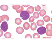 |
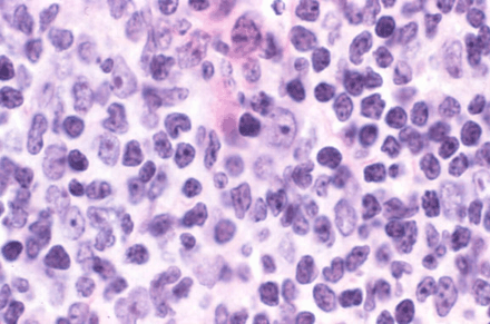
| Hematoxylin and Eosin (H & E) smear from FL tissue. | Example morphology of a FL in a peripheral blood. Note cleaved nuclei. |
Immunophenotyping
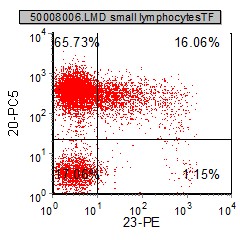
When using fresh tissue for flow cytometric immunophenotyping, the predominant populations are lymphoid. these can be stratified as large and small lymphocytes (CD45 positive). The gating dot plot below identifies a predominant CD45 bright, FS (small used cells). These cells were in the subsequent anlysis.
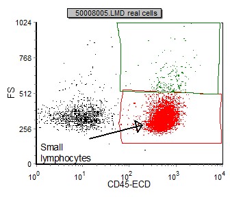 |
| Example of a gating dot plot for a tissue sample CD45 vs FS. The predominant red population is the small lymphoid cells used for further analysis. |
Below is a table of common immunophenotyping markers used to identify and classify Follicular Lymphoma (FL).
| Marker | Expression in FL |
| CD5 | - |
| CD10 | +/- |
| CD11c | -/+ |
| CD19 | + (dim) |
| CD20 | + (moderate) |
| CD22 | + (moderate) |
| CD23 | +/- |
| CD25 | - |
| CD30 | - |
| CD38 | +/rare - |
| CD43 | -/rare + |
| CD45 | + |
| CD103 | - |
| CD138 | - |
| bcl-1 | - |
| bcl-2 | + |
| bcl-6 | + |
| light chain (kappa or lambda) | +(occassional sIg -) |
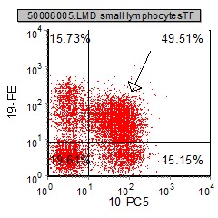
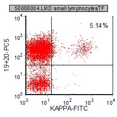
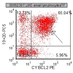
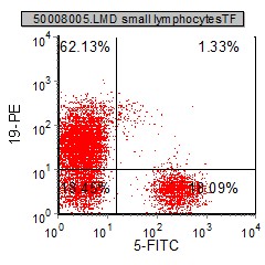
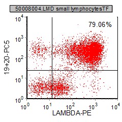
Below are selected example dot plots from a case of FL:
 |
 |
 |
 |
 |
 |
| FL characteristically coexpresses CD19 and CD10. CD5 is absent on the B cells. | Above is an example of the monoclonal lambda light chain restriction in this case. sIg expression is moderate to bright. | cytoplasmic bcl-2 is characteristically positive on the B cells. CD23 is absent and CD20 is positive and generally more positive than CD19. |
Other relevant tests
Cytochemistry: OCT 2, BOB 1, Ki-67 and PAX 5 by immunohistochemistry are expressed on FL cells.
Genetics: A translocation between chromosome 14 and 18 results in the overexpression of the bcl2 gene. This overexpression causes a blockage of apoptosis, or programmed cell death. This translocation has been associated with the development of Follicular lymphoma.
Flow Diagnosis
FL cells are surface Ig+ (heavy and light chain - predominantly IgM). they express bcl-2, CD10, CD19, CD20, CD22 and CD79a. CD5 and CD23 are characteristically negative. Surface light chains (kappa or lambda) and CD20 are distinctly brighter thanCLL/SLL.