Plasma Cell Dyscrasias (PCD)
You are here
Definition
Plasma cell dyscrasias (include multiple myeloma) are a multifocal clonal plasma cell proliferation infiltrating the bone marrow. This disease can be clinically indolent or aggressive.
Sample Cases
Click here for instructions on how to download the free FCS Express Reader to view and manipulate the sample cases.
| Case Name (click on case name to open) |
Comments | Size |
| PCD1 |
An interesting case with a differential of Hairy Cell Leukemia or PCD. See how to make the call. |
9 Mb |
| PCD2 |
PCD |
5 Mb |
| PCD3 |
PCD with dim/mod CD45 and dim CD19 expression. |
5 Mb |
| case 36 | Waldenstrom's Macroglobulenemia This case was kindly provided by the ASCP Press. It is part of Flow Cytometry in Clinical Diagnosis by John Carey, Phil McCoy and David Keren. |
18.2 MB |
| case 35 | CD56+ clonal PCD This case was kindly provided by the ASCP Press. It is part of Flow Cytometry in Clinical Diagnosis by John Carey, Phil McCoy and David Keren. |
2.65 MB |
| 38+56+19-PCD.fey | A PCD case showing dim/neg CD45 cells co-expressing CD38, CD56 and CD19-. | 6.56 MB |
Epidemiology
Plasma cell myeloma is the most common lymphoid malignancy in Blacks and the second most common in Whites. It represents 15% of hematologic malignacies in the United States. The median age at diagnosis is 68 years of age for males and 70 for females, and there is a 1:1 male to female ratio.
Possible causes
The higher incidence in Blacks mirrors their high immunoglobulin level, which suggests a greater B cell population. There is an increased risk in farmers and cosmetologists. Specific exposure includes pesticides, petroleum, asbestos and rubber along with high doses of radiation. A possible link to viruses is noted in the literature; specifically HIV and HHV8.
Morphology
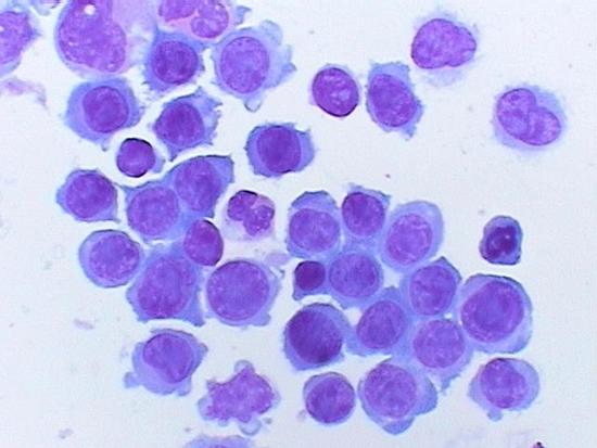
Mature plasma cells are usually oval with a round eccentric nuclei. There is abundant basophillic cytoplasm and a marked perinuclear hof. Plasmablasts are multinucleated and polylobulated. (examples below)
 |
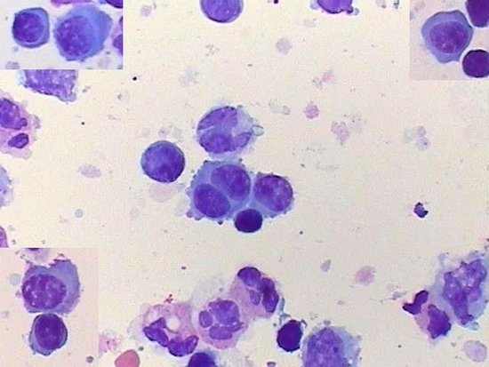 |
| Example morphology of plasma cells. | Example morphology of plasma cells. |
Immunophenotyping
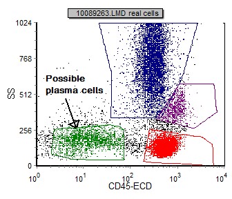
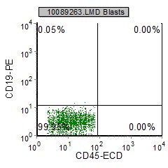
Plasma cells can be initially identified using a SSC vs CD45 plot. These cells have low to moderate SSC and negative CD45. (Occasionally plasma cells will be dim or moderate CD45). A population of > 0.5% that is negative CD45 and brightly positive for CD38 (4th log decade) is highly suspicious of plasma cells and should be further investigated.
 |
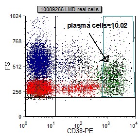 |
| Plasma cells are generally CD45 negative and low to moderate SSC. Other cells may fall in this region such as red cell precursors etc. | Plasma cells express very bright CD38 (usually in the 4th decade). When a population of bright CD38 is present >1% it should be further investigated. |
Once the plasma cell gate is established, the following markers are useful in subclassification of PCD:
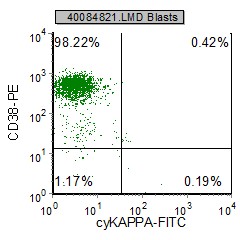
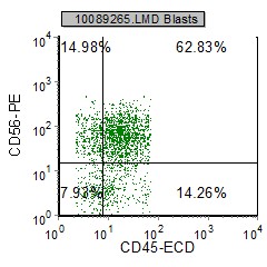
Plasma cells are positive for CD38 (brightly expressed), CD138, cytoplasmic light (either kappa or lambda) and heavy chain immunoglobulins. Surface immunoglobulins (kappa and lambda) are usually negative; however, posive for cytoplasmic expression. IgG is the most common heavy chain followed by IgA. More than 50% of the plasma cell neoplasms express CD117 and or CD56, along with CD43. Myeloma cells usually do not express CD45, but can be dim and occassionally moderate. CD45 is also shown to correlate with the proliferation rate of the myeloma cells. (S-phase fraction or Ki-67)
| Marker | % expressed in PCD |
| CD19 | 6.5 |
| CD20 | 30.9 |
| CD38 | 98.4 |
| CD138 | 96.2 |
| CD43 | 51.5 |
| CD45 | 21.4 |
| CD56 | 53.8 |
| CD79a | 26.7 |
| CD117 | 63.2 |
| bcl-1 | 34.1 |
| bcl-2 | 53.3 |
| IgM | 56.4 |
| IgA | 20 |
| IgM | 5.5 |
| IgD | 1.8 |
| heavy chains absent | 11 |
| cyKappa | 57.3 |
| cyLambda | 41.2 |
| absent light chains | 1.5 |
Example histograms in PCD
 |
 |
 |
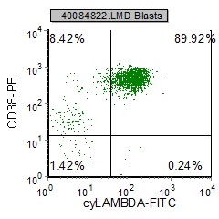 |
| Plasma cells are CD45 and CD19 negative. Neoplastic plasma cells frequently express CD56. | Neoplastic plasma cells do not express surface light chains; however, cytoplasmic monoclonal light chains are expressed in plasma cell neoplasms. (Above is a cytoplasmic lambda monoclonality) |
Other relevant tests
Cytochemistry: MUM-1, OCT-2 and BOB-1 are expressed by immunohistochemistry on PCD.
Genetics: MM with t(11:14)(q(13;q32) can display upregulation of bcl-1/cyclin D1. High proliferations can be associated with deletions of 13q14 and 17p13.
Flow Diagnosis
To diagnose neoplastic plasma cells. A population of CD45 and CD19 negative cells with bright CD38 and CD138. These cells express a cytoplasmic light chain expansion (either kappa or lambda). CD56 and/or CD117 can also be positive.
References
Rawston AC. Immunophenotyping of Plasma Cells, Current Protocols in Cytometry Unit 6.23, May 2006