Acute Minimally Differentiated Leukemia (M0)
You are here
Please contribute to the site. Contact us for more information.
Definition
Myeloid Leukemia with Minimal Differentiation - FAB M0. M0 is an acute leukemia with no evidence of myeloid differentiation by light mocroscopy. Immunophenotyping studies are essential in all cases to distinguish this disease from acute lymphoblastic leukemia (ALL).
Sample Cases
Click here for instructions on how to download the free FCS Express Reader to view and manipulate the sample cases.
| Case Name (click on case name to open) |
Comments | Size |
| M0-1 | Sample AML case (M0 Subtype) | 3.1 Mb |
| Case 34 | M0 AML This case was kindly provided by the ASCP Press. It is part of Flow Cytometry in Clinical Diagnosis by John Carey, Phil McCoy and David Keren. |
1.47 MB |
Epidemiology
These cases comprise approximately 5% of cases of AML Most cases are seen in adults.
Possible causes
Unknown
Morphology
The blasts are of medium size with dispersed nuclear chromatin, round or indented nuclei with one or more nucleoli. The cytoplasm is agranular with varying degrees of basophila.
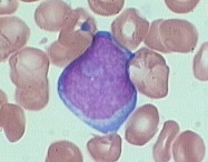 |
| Example morphology of AML M0 blast. |
Immunophenotyping
Below is the CD45 vs SSC gating plot. M0 blasts are CD45 dim and SSC low (green). They are a similar size to normal lymphocytes (red).
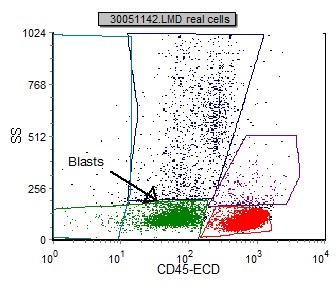 |
| Example of M0 blasts in a CD45 vs SSC dot plot. |
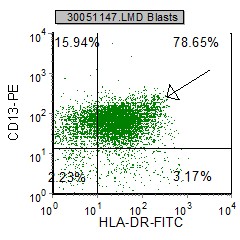
M0 blasts are negative for B and T lymphoid antigens, platelet glycoproteins GP Ib and GP IIb/IIIa, and erythroid glycophorin A. Myeloid antigens such as CD13, CD33 and CD11b are variably positive. CD34 and HLA-DR are generally positive. MPO is negative for these blasts. Below are selected histograms from an M0 case.
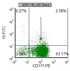 |
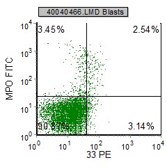 |
 |
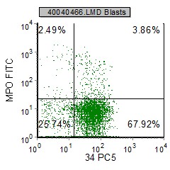 |
| HLA Dr, CD13 and CD117 are expressed. CD15 is absent. | CD34 is expressed, CD33 is absent and MPO is minimally expressed (~5%) |
Other relevant tests
Cytochemistry: M0 blasts are nondescript (no Auer rods) and are MPO, PAS and NSE negative (<3%). Electron microscopic ultracytochemical studies for platelet peroxidase are negative.
Genetics: There is no particular association with any chromosome abnormalities.
Sub-classification
None
Flow Diagnosis
These blasts are CD45 dim and express the primitive hematopoetic markers CD34, CD38 and HLA Dr. TdT may be expressed in 1/3 of cases. One or more of the following pan myeloid markers are expressed; CD13, CD33 and/or CD117. B and T restricted antibodies are not expressed (cCD3, cCD79a, cCD22). MPO is negative on these cases, though very few blasts may express MPO. Occassionally CD2 and/or CD7 (not lymphoid specific) are expressed on these cells.