Acute Myelogenous Leukemia (AML) not otherwise categorized - General Information
You are here
Definition
Acute myelogenous leukemia (AML) is a clonal expansion of the myeloid blasts in bone marrow, blood or other tissues.
Epidemiology
The incidence of acute leukemia is approximately 4 cases per 100,000 per year. 70% of these are AML. AML is generally seen in adults with a median age of 60 years. In this age group the incidence is 10 cases per 100,000. There are approximately 9700 cases of AML in the United States per year. The male female ratio for AML is 1:1.
Subclassification and frequency of AML
| Subclassification | Frequency |
|
Acute nonlymphocytic (ANLL) |
|
|
M1 Myeloblastic (AML) |
10-20% |
|
30-40% |
|
|
10-15% |
|
|
M4 Myelomonocytic(AMML, Naegeli) |
10-15% |
|
M5a Monoblastic (AMoL, Schilling) |
10-15% |
|
<5% |
|
|
M6 Erythroleukemia (Di Guglielmo) |
<5% |
|
<5% |
|
|
Other (e.g. biphenotypic) |
<5% |
Possible causes
Similar to all, possible factors that have been associated with AML (and MDS) are viruses, radiation, cytotoxic chemotherapy and benzene exposure. Exposure to the atomic bomb of WWII increased the incidence of all leukemias including AML. In addition, smoking is attributed to an increased risk.
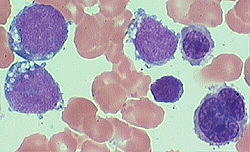
Morphology
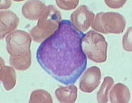
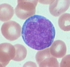
Myeloblasts vary in size from slightly larger than lymphocytes to larger than a monocyte. These blasts have abundant basophillic cytoplasm. The nucleoi are round with fine chromatin and multiple nucleoli. Some azurophillc granules or Auer rods are present in the cytoplasm.
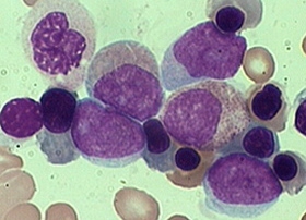
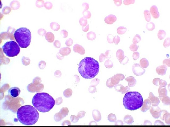
Below are example morphology of selected AML types:
 |
 |
 |
| Example morphology of AML-M0 | Example morphology of AML-M1 | Example morphology of AML-M2 |
 |
 |
 |
| Example morphology of AML-M3 (hypercellular) | Example morphology of AML-M5 | Example morphology of AML-M6 |
Sub-classification of AML
The WHO classification for AML is:
1. Acute myeloid leukemia with recurrent genetic abnormalities
- Acute myeloid leukemia with t(8;21)(q22;;q22); AML1/ETO
- Acute myeloid leukemia with abnormal bone marrow eosinophilia inv(16)(p13q22) or t(16;16)(p13;q22): CBFB/MYH11)
- Acute Promyelocytic Leukemia - AML with t(15;17)(q22;q12); PML/RARa and variants (M3)*
- Acute myeloid leukemia with 11q23 (MLL) abnormalities.
2. Acute myeloid leukemia with multilineage dysplasia
3. Acute myeloid leukemia and myelodysplastic syndrome, therapy related
4. Acute myeloid leukemia not otherwise categorized
-
Acute basophillic leukemia
-
Acute panmyelosis with myelofibrosis
-
Myeloid sarcoma
(flow cytometric immunophenotyping is helpful in diagnosis and/or classification) *
The 2006 International Bethesda Consensus recommends the following CD markers for the initial evaluation of myeloid leukemias:
CD7, CD11b, CD13, CD14, CD15, CD16, CD33, CD34, CD45, CD56, CD117, HLA Dr.
Additional CD Markers for secondary evaluation of myeloid lineage are:
CD2, cCD3, CD4, cCD22, CD25, CD36, CD38, CD41, CD61, cCD61, CD64, CD71, cCD79a, cMPO, CD123, CD163, CD235a.
Immunophenotyping
The following is a table that indicates the specific immunophenotypic markers and their reactions for each type of AML:
| Marker | AML-M0 | AML-M1 | AML-M3 (APL) | AML-M4 (blasts) | AML-M4 (monos) | AML-M5 | AML-M6 | AML-M7 |
| CD2 | - | - | +/- | - | - | - | - | - |
| CD3 | - | - | - | - | - | - | - | - |
| CD4 | +/- | +/- | +/- | +/- | + | + | - | -/+ |
| CD7 | -/+ | -/+ | -/+ | -/+ | +/- | +/- | - | -/+ |
| CD10 | - | - | - | - | - | - | - | - |
| CD11b | - | - | - | - | + | + | - | - |
| CD11c | -/+ | -/+ | -/+ | +/- | + | + | - | - |
| CD13 | + | + | + | + | + | dim+ | - | -/dim+ |
| CD14 | - | - | - | - | + | +/- | - | - |
| CD16 | - | - | - | - | - | -/+ | - | - |
| CD19 | - | + | - | - | - | - | - | - |
| CD20 | - | - | - | - | - | - | - | - |
| CD22 | - | - | - | - | - | - | - | - |
| CD33 | + | + | + | + | + | bright+ | - | bright+ |
| CD34 | + | + | - | + | - | +/- | +/- | -/rare+ |
| CD41 | - | - | - | - | - | - | - | + |
| CD45 | + | + | + | + | + | + | -/rare+ | +/rare- |
| CD56 | -/+ | +/rare- | -/+ | - | -/+ | +/- | -/rare+ | -/rare+ |
| CD61 | - | - | - | - | - | - | - | + |
| CD64 | -/+ | -/+ | -/+ | -/+ | + | +/- | - | - |
| CD79a | - | - | - | - | - | - | - | - |
| CD117 | + | + | + | + | - | -/+ | dim+ | dim+ |
| HLA-DR | +/rare- | +/rare- | - | + | + | +/- | + | -/dim |
| TdT | -/+ | -/+ | - | - | - | - | - | - |
| GPHA | - | - | - | - | - | - | + | - |
| cIgM | - | - | - | - | - | - | - | - |
Flow Diagnosis
To diagnose, please see the specific AML type below
- Acute Erythroblastic leukemia (M6)
- Acute Megakaryoblastic Leukemia (AMKL) (M7)
- Acute Minimally Differentiated Leukemia (M0)
- AML (Acute Myelogenous Leukemia with maturation) (M2)
- AML (Acute Myelogenous Leukemia without maturation) (M1)
- AMML (Acute Myelomonocytic Leukemia) (M4)
- AMoL (Acute Monoblastic/Monocytic Leukemia) (M5)