AML (Acute Myelogenous Leukemia without maturation) (M1)
You are here
Definition
Acute Myeloblastic Leukemia without maturation - FAB M1: AML-M1 is defined and characterized by a high percentage of blasts in the bone marrow without significant evidence of myeloid maturation. Blasts constitute >90% of the nonerythroid cells. The myeloid nature of the blasts is demonstrated by MPO or SBB (>3% of blasts) positivity and/or Auer rods.
Sample Cases
Click here for instructions on how to download the free FCS Express Reader to view and manipulate the sample cases.
| Case Name (click on case name to open) |
Comments | Size |
| case 20 | AML M1 This case was kindly provided by the ASCP Press. It is part of Flow Cytometry in Clinical Diagnosis by John Carey, Phil McCoy and David Keren. |
2.06 MB |
Epidemiology
AML-M1 comprises approximately 10% of AMLs. It may occur at any age but the majority of patients are adults. The median age is 46 years of age.
Possible causes
Unknown.
Morphology
In some cases the immature cells have abundant, frequently basophilic cytoplasm, with variable numbers of often indistinct, sometimes coalescent granules. If such immature cells are < 10% the diagnosis is M1, but if > 10% the diagnosis becomes
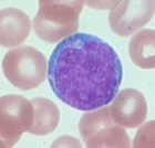 |
| Typical morphology of AML-M1 blast. |
Immunophenotyping
Using a CD45 vs SSC gating dot plot, blasts can be identified (green population below). Myeloblasts M1 have moderate CD45 expression and low to moderate SSC. Note these blasts have a slightly higher SSC than the lymphocytes (red population below).
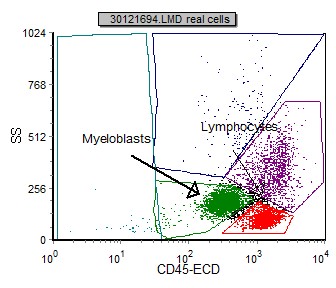 |
| An example CD45 vs SSC gating dot plot identifies myeloblasts in a case of AML-M1. |
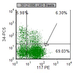
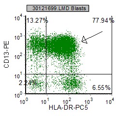
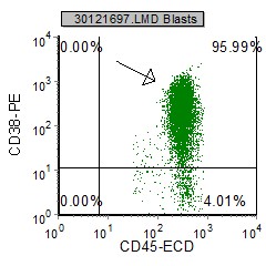
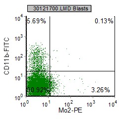
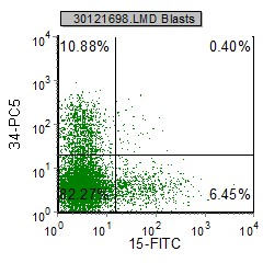
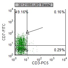
Below are selected example histograms from a case of AML M1.
 |
 |
 |
 |
 |
 |
| CD34, CD11b and Mo2 are negative on the blasts. CD117 is expressed on these blasts. |
HLA Dr and CD13 are expressed on the blasts, where CD34 and CD15 are not. |
CD38 is positive, and CD7 is partially expressed on these blasts. |
The M1 blasts express at least two of the following myeloid antigens: CD13, CD33, CD117, MPO and/or HLA-DR. CD34 is often positive. There is generally no expression of the monocytoid markers CD11b or CD14. Lymphoid antigens CD3, CD20, CD79a are absent. CD7 may be expressed.
Other relevant tests
Cytochemistry: Relatively few blasts (5-10%) are MPO (myeloperoxidase) positive. A minimum of 3% MPO positive blasts are required for diagnosis. NSE and PAS are generally negative.
Genetics: Chromosome Abnormalities: t(9;22) Philadelphia chromosome, 8+, -5, and -7.
Flow Diagnosis
AML-M1 and AML-M2 are initially stratified by morphology (see above). M1 blasts must express at least two of the following myeloid antigens: CD13, CD33, CD117, MPO and/or HLA-DR. MPO must be expressed on > 3% on blasts.