AMML (Acute Myelomonocytic Leukemia) (M4)
You are here
Definition
Acute Myelomonocytic Leukemia (AMML) - AML-M4 is defined as an acute leukemia with differentiation along both myeloid and monocytic lines. Monocytes and promonocytes represent > 20%, but < 80% of the marrow differential. Both myeloblasts and monoblasts are present. A high number of circulating monocytes may be present.
Sample Cases
Click here for instructions on how to download the free FCS Express Reader to view and manipulate the sample cases.
| Case Name (click on case name to open) |
Comments | Size |
| M4-1 | M5b or M4 | 8.5 Mb |
| Case 2 | AMoML case submitted by UTMC | 2.6 Mb |
Epidemiology
Acute myelomonocytic leukemia comprises approximately 15-25% of all cases of AML. Some patients have a history of CMML. It occurs in all age groups, but is more common in older adults. The median age is 50 years of age. The male to female ratio is 1.4 to 1.
Possible causes
Unknown.
Morphology
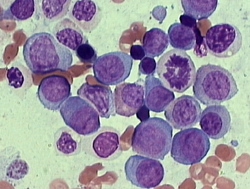
Monoblasts are large cells with abundant cytoplasm which can be moderately to intensely basophillic. There may be scattered azurophillic granuoles and vacuoles. The monoblasts will have lacy nuclear chromatin and one or more predominant nucleoli. A variant of AML-M4 has an increase in eosinophils and is classified as AML-M4e. Typical morphology is displayed below.
 |
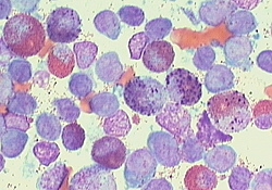 |
| Morphology of M4 AML | Morphology of M4e AML |
Immunophenotyping
The CD45 vs SSC gating dot plot in a case of AML-M4 shows a population of CD45 moderate, SSC low myeloblasts and a population of monocytoid cells, which include monoblasts (CD45 bright and SSC low-moderate)
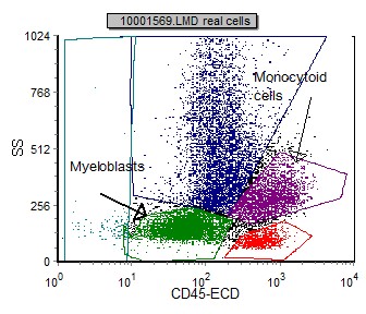 |
| Typical CD45 vs SSC gating dot plot of AML-M4 |
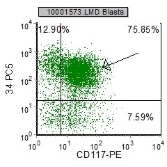
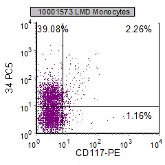
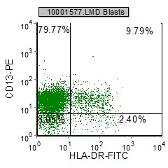
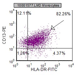
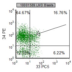
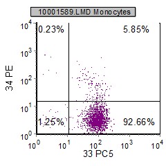
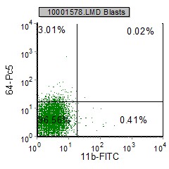
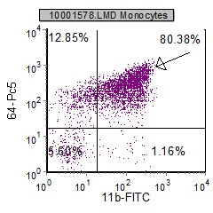
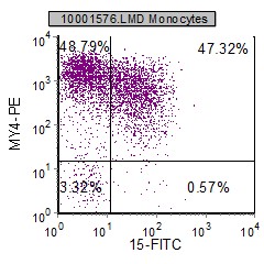
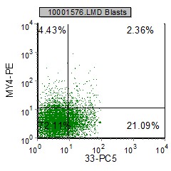
Below is a comparison of selected markers on this case of both the myeloblasts and monocytoid cells used to differentiate AML-M4. The myeloblasts marker expression is in the left column (green), and the monocytoid cells marker expression is in the right colum (purple).
 |
 |
 |
 |
 |
 |
 |
 |
 |
 |
 |
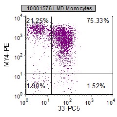 |
In general, myeloblasts (green) are moderate CD45 and low SSC and express CD13, CD33, CD34, CD117, HLA Dr. Monocytoid cells (purple) including monoblasts and promonocytes are CD45 bright and SSC slightly higher than myeloblasts. These cells express CD11b, CD11c, CD13, CD14, CD33, CD64 and HLA Dr.
Other relevant tests
Cytochemistry: CAE, usually negative in eosinophils, is frequently positive in the abnormal eosinophils of M4e. More than 20% of the blasts should be MPO + and more than 20% should be NSE + .
Genetics: Chromosome abnormalities: t(4;11), t(9;11), 8+ and -7. Variant: M4e variant in which eosinophils (> 5%) are increased in number and abnormal associated with abnormalities of chromosome 16.
Diagnosis Aides:
- High serum lysozyme (3x normal)
- A peripheral monocytosis of > 5x10/L in an otherwise M2 marrow and increased lysozyme
- A peripheral monocytosis of >5 x10/L in M2 marrow and >20% NSE + marrow blasts.
Sub-classification
Morphologically their are two types of AML-M4 based on morphology. AML-M4 and AML-M4e variant which has an increased eosinophil count.
Flow Diagnosis
The diagnosis of AML-M4 initially needs a morphologic review and categorization. AML-M4 cases must have a population of myeloblasts and monoblasts (>20%). Monoblasts must be < 80% of total nucleated cells. The flow immunophenotype will confirm the gated cell types and thus confirm the diagnosis. In general, myeloblasts (green) are moderate CD45 and low SSC and express CD13, CD33, CD34, CD117, HLA Dr. Monocytoid cells (purple) including monoblasts and promonocytes areCD45 bright and SSC slightly higher than myeloblasts. These cells express CD11b, CD11c, CD13, CD14, CD33, CD64 andHLA Dr.