AMoL (Acute Monoblastic/Monocytic Leukemia) (M5)
You are here
Definition
Acute monocytic leukemia (AMoL, or AML-M5) is considered a type of acute myeloid leukemia (AML). In order to fulfill World Health Organization (WHO) criteria for AML-M5, a patient must have greater than 20% blasts in the bone marrow, and of these, greater than 80% must be of the monocytic lineage. A further subclassification (M5a versus M5b) is made depending on whether the monocytic cells are predominantly monoblasts (>80%) (acute monoblastic leukemia) or a mixture of monoblasts and promonocytes (<80% blasts).
Sample Cases
Click here for instructions on how to download the free FCS Express Reader to view and manipulate the sample cases.
| Case Name (click on case name to open) |
Comments | Size |
| M5_1 | M5 AML | 5.59 MB |
| M5_2 | AML M5b | 7.74MB |
Epidemiology
Acute monoblastic leukemia comprises 5-8% of cases of AML. It may occur at any age but is most common in young individuals. In infancy, it is frequently associated with abnormalities of 11q23. Extramedullary lesions may occur. Acute monocytic leukemia conmprises 3-6% of cases; the male to female ratio is 1.8 to 1.0. It is more common in adults. The median age is 49 years.
Possible causes
None known.
Morphology
Monoblasts can be distinguished by having a roughly circular nucleus, delicate lacy chromatin, and abundant, often basophilic cytoplasm. These cells may also have pseudopods. By contrast, promonocytes have a more convoluted nucleus, and their cytoplasm may contain metachromatic granules.
 |
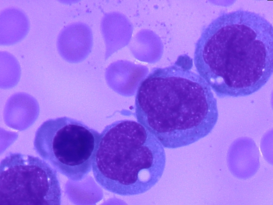 |
| Example morphology of acute monocyticleukemia case. There is an admix of monoblasts and monocytes. (note granules) | Example morphology of an acutemonoblastic leukemia case. Monoblasts predominate. |
Immunophenotyping
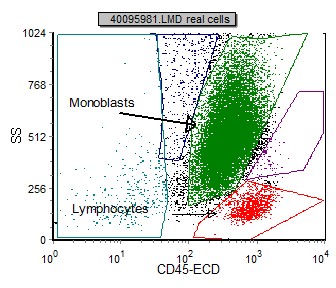
Neoplastic cells in AMoL can be initially identified using a SSC vs CD45 plot. Depending on the maturation of the cells of interest, they will fall into a characteristic region on the gating dot plot. Monoblasts have little to no granuoles and therefore have a lower side scatter than a monocytic leukemia which have a higher and more variable SSC. These cells may also have different fluorescent intensity of CD45. More mature monocytoid cells will have a brighter CD45 and fall closer to the monocyte region. See examples below.
 |
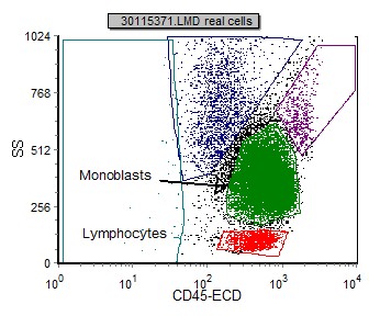 |
| This CD45 vs SSC plot corresponds to the above acute monocytic leukemia case. Abnormal monocytic population is green. Note the higher SSC. |
This CD45 vs SSC plot corrponds to the above acute monoblastic leukemia case. Abnormal monocytic population is green. Note the lower SSC. |
Below is a list of immunophenotypic markers that are useful in the identification and classification of AMoL:
|
Marker |
Prevalence in AMoL |
| CD2 | 10% |
| CD4 | 93% |
| CD7 | 11% |
| CD10 | 11% |
| CD11b | 85% |
| CD11c | 100% |
| CD13 | 77% |
| CD14 | 54% |
| CD16 | 13% |
| CD23 | 30% |
| CD33 | 100% (bright) |
| CD34 | 3% |
| CD45 | 100% (mod-bright) |
| CD56 | 80% |
| CD64 | 96% |
| CD117 | 14% |
| HLA Dr | 93% |
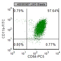
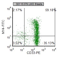
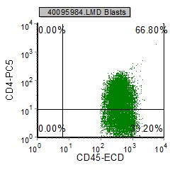
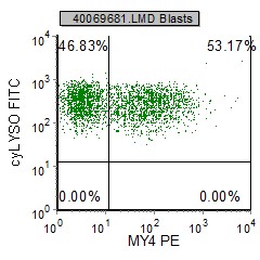
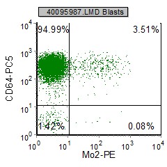
Below are selected example dot plots from a AMoL case:
 |
 |
 |
 |
 |
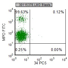 |
| CD64, CD11b and Lysosome are characteristically positive in AMoL. My4 can be partially expressed depending on the stage of maturity of the cells. (In this case My4 is variably expressed) | CD33 and CD64 are consistently expressed. (CD33 is generally bright). My4 and Mo2 are variably expressed depending on maturity of cells. | In this case CD4 is dimly expressed on these cells. CD34 is characteristically negative. Note the heterogenous population of MPO indicating a variety of maturity level of the cells in question. |
Sub-classification
A further sub-classification (M5a versus M5b) is made depending on whether the monocytic cells are predominantly monoblasts (>80%) (acute monoblastic leukemia) or a mixture of monoblasts and promonocytes (<80% blasts).
Other relevant tests
Cytochemistry: Monoblasts are typically MPO negative and promonocytes are MPO variable. Both monoblasts and promonocytes stain positive for non-specific esterase (NSE), however NSE may often be negative.
Genetics: There is a strong association with AMoL and deletions and translocations involving chromosome 11 band 23. Translocation t(8:16)(p11;p13)may be associated with AMoL and AMML.
Flow Diagnosis
Acute monoblastic/monocytic leukemia (M5) blasts express characteristically express CD4, CD11b, CD11c, CD13 (dim), CD33(bright), CD45 (bright-mod), CD56, CD64 and HLA Dr. A subset of these cases may also express CD2, CD7, CD10, CD16, CD23, lysosome and CD117. CD34 is predominantly negative. Though normal monocytes express CD14, in AMoL, this marker is variably expressed (predominantly negative).